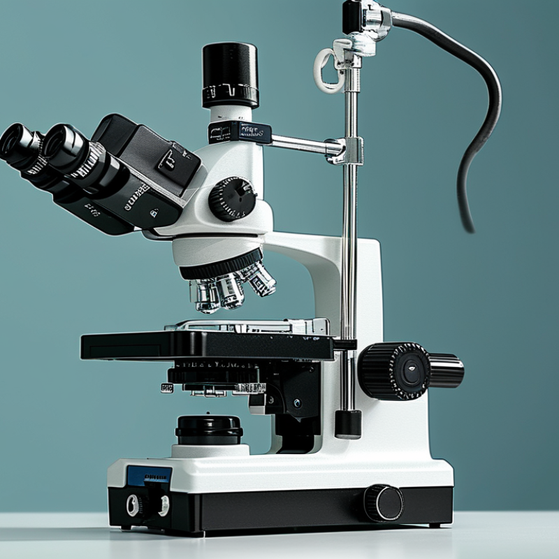
| Optical Quality | Magnification range | A continuous or multi-step range covering approximately 3× to 20×, with clearly specified values for each position. | Low magnification supports orientation and setup; higher magnification helps inspect the tympanic membrane, ossicles, laryngeal structures, and fine anatomy. | Review the optical specification sheet and test the full range on a calibrated target and representative anatomical models. | High | 10% |
| Optical resolution | Resolution should be stated using a defined test method, with no visible loss of fine detail across the central field and clinically relevant magnifications. | Higher resolving power can improve identification of small anatomical boundaries and tissue details. | Request measured resolution data, test a USAF 1951 resolution target, and compare images at the same working distance and illumination level. | High | 9% |
| Contrast and color fidelity | Neutral color rendering, controlled chromatic aberration, and sufficient contrast without excessive artificial sharpening or color cast. | Accurate color and contrast assist assessment of inflammation, vascularity, mucosal changes, and tissue margins. | Use a color chart, grayscale chart, and low-contrast target under clinical illumination; compare optical viewing with recorded output. | High | 8% |
| Depth of field and focus stability | Stable focus during small movements, with smooth fine focusing and no noticeable focus drift after the system reaches operating temperature. | ENT procedures often involve curved or uneven anatomy and frequent instrument movement. | Test on stepped-depth targets and during simulated suction, drilling, and instrument manipulation. | High | 7% |
| Illumination uniformity | A bright, centered, shadow-controlled field with no obvious hot spot, edge darkening, or flicker at normal working distances. | Uniform illumination reduces the need for repeated repositioning and helps reveal anatomy in narrow, deep spaces. | Measure illuminance at the center and edges of the field using a calibrated light meter; inspect the field with and without instruments present. | High | 8% |
| Working distance | A practical working distance commonly around 200–400 mm, with the selected configuration matched to the surgeon’s posture and instruments. | The correct distance affects access, instrument clearance, surgeon posture, and compatibility with sterile drapes. | Confirm the working distance from the manufacturer’s technical drawing and conduct a hands-on procedure simulation. | High | 6% |
| Ergonomics and Handling | Binocular head and interpupillary adjustment | Smooth independent adjustment with a broad interpupillary range and secure locking without image shift. | Correct alignment reduces eye strain and helps multiple users share the microscope efficiently. | Have users with different interpupillary distances perform repeated setup and adjustment cycles. | High | 6% |
| Eyepiece and viewing angle | Ergonomic eyepieces or an adjustable observation system that supports neutral neck and shoulder posture during long procedures. | Awkward viewing angles can contribute to fatigue, neck flexion, and reduced concentration. | Assess posture during a 30–60 minute simulated procedure using the actual operating chair, table, and instruments. | High | 7% |
| Stand movement and balance | Smooth, controlled movement in all required axes, with reliable brakes or electromagnetic locks and minimal drift after positioning. | Precise positioning is essential when working around delicate ENT structures. | Move the microscope through the full range with accessories attached; assess drift, vibration, brake response, and collision clearance. | High | 7% |
| Control accessibility | Frequently used functions, including focus, magnification, illumination, and recording, should be reachable without breaking the sterile workflow. | Accessible controls reduce interruptions and unnecessary hand or body movement. | Use sterile covers and evaluate footswitches, hand controls, and programmable buttons during a simulated case. | Medium | 5% |
| Equipment footprint and integration | The stand, monitor, accessories, and cable routing should fit the operating room without blocking anesthesia access, staff circulation, or emergency movement. | Space constraints can affect workflow and safety more than optical specifications alone. | Review a scaled room layout and perform an on-site clearance check with all connected equipment installed. | Medium | 4% |
| Imaging and Documentation | Camera resolution and sensor performance | A medical-grade camera with documented resolution, adequate low-light performance, and a sensor format appropriate for the optical path. | Image quality must remain useful when illumination is reduced or when the anatomy contains both bright and shadowed areas. | Record standardized targets at several magnifications and illumination levels; inspect fine detail, noise, and highlight clipping. | High | 8% |
| Video format and frame rate | At least Full HD for routine documentation; 4K may be appropriate where large displays, teaching, or detailed archiving justify the additional storage and workflow requirements. | Higher resolution can support teaching and review, while adequate frame rate helps display smooth instrument movement. | Confirm supported resolution, frame rate, latency, file format, and compatibility with the hospital display and recording system. | High | 6% |
| Image synchronization | The recorded image should remain synchronized with the live optical view and should not show clinically distracting latency or dropped frames. | Synchronization is important for teaching, remote consultation, and coordinated instrument handling. | Record a moving target and compare live and recorded output; test the complete signal path rather than the camera alone. | Medium | 4% |
| Connectivity and interoperability | Documented outputs and protocols suitable for the facility’s monitors, capture devices, network environment, and electronic records workflow. | Open, documented connectivity reduces dependence on proprietary accessories and simplifies future upgrades. | Verify connector types, output resolutions, network requirements, export formats, and integration responsibilities in writing. | High | 5% |
| Data security and patient privacy | Configurable user access, controlled export, secure storage options, and a documented process for handling patient-identifiable images. | Recorded endoscopic images may contain protected health information and require controlled access. | Review the system’s user-management, audit, storage, deletion, and export procedures with the hospital’s IT and compliance teams. | High | 4% |
| Safety, Compliance, and Reliability | Electrical and electromagnetic safety | Applicable medical electrical equipment documentation, including conformity to relevant electrical safety and electromagnetic compatibility requirements for the target market. | Compliance supports safe operation around patients and other electrically sensitive equipment. | Request current declarations, test reports, certificates, labeling information, and country-specific regulatory documentation. IEC 60601-1 and IEC 60601-1-2 are common reference standards. | Critical | 7% |
| Risk management and usability evidence | A documented medical-device risk-management process and usability engineering evidence appropriate to the device and intended use. | Formal risk analysis helps address foreseeable misuse, positioning hazards, electrical risks, and workflow-related errors. | Review the applicable risk-management summary and usability documentation. ISO 14971 is a common reference for medical-device risk management. | Critical | 5% |
| Reliability and serviceability | Documented preventive-maintenance intervals, replaceable wear components, spare-parts availability, and a defined repair escalation process. | Downtime can disrupt scheduled surgery and may create costly emergency replacements. | Request service-level terms, maintenance schedules, mean-time or failure data when available, and references from comparable clinical installations. | High | 5% |
| Cleaning, disinfection, and sterile compatibility | Clear instructions for cleaning and disinfection, compatible sterile drapes, sealed control surfaces where appropriate, and materials resistant to approved agents. | ENT environments require repeatable infection-control procedures without damaging optical or electronic components. | Compare the instructions for use with the facility’s approved disinfectants and observe a complete setup and turnover cycle. | Critical | 4% |
| Supplier Capability | Application support and training | Structured installation, user training, clinical application support, and refresher training for surgeons, nurses, technicians, and biomedical staff. | Correct setup and operation directly affect image quality, ergonomics, and equipment lifespan. | Evaluate the training agenda, trainer qualifications, competency records, and post-installation support process. | High | 4% |
| Warranty and total cost of ownership | A transparent five-to-seven-year ownership model including purchase price, installation, service, consumables, accessories, software, training, and expected upgrades. | The lowest purchase price may not represent the lowest operational cost. | Request a line-item quotation and calculate annualized ownership cost under realistic service and accessory assumptions. | High | 5% |
| Evidence-based acceptance testing | The supplier should agree to measurable site-acceptance criteria covering optics, illumination, movement, imaging, safety documentation, and user training. | Acceptance testing converts general marketing statements into verifiable procurement requirements. | Include pass/fail criteria, test equipment, responsible parties, remediation timelines, and final sign-off in the purchase contract. | Critical | 4% |